Scientists analyzed individual cells isolated from two craniopharyngioma subtypes to identify the specific cell types, their features, and their crosstalk
Craniopharyngiomas are brain tumors that negatively impact the hormonal function of the nearby pituitary. The tumor location often prevents necessary surgical intervention. Alternative pharmacological therapy requires an in-depth understanding of the tumor molecular characteristics. To address this gap, researchers from Japan analyzed gene expression within individual tumor cells. This study reports the molecular features and interactions of tumor and immune cells associated with two craniopharyngioma subtypes that will help identify future targeted therapeutics.

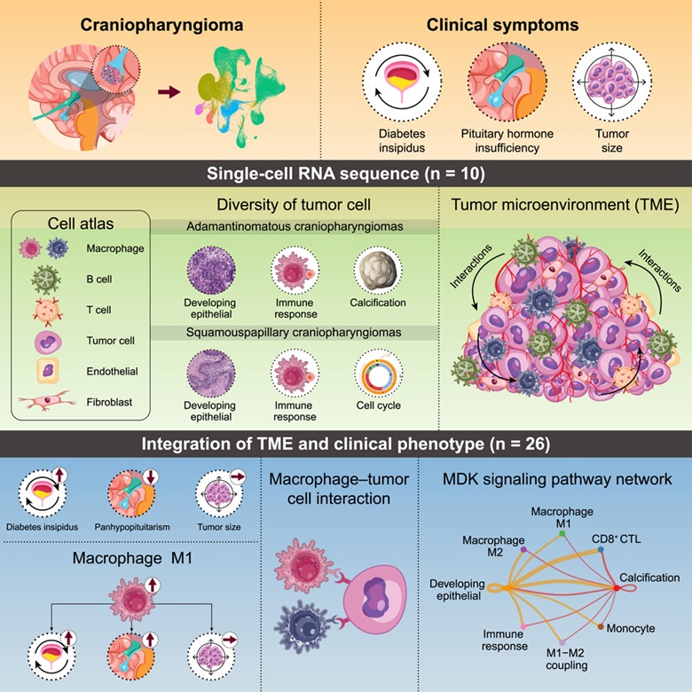
Image title: Craniopharyngioma, a rare and benign tumor is challenging to treat because of its proximity to the pituitary gland.
Image caption: The study explored the complex interactions between tumor cells and immune cells, including T cells, M1/M2 macrophages, and their localized impact on the tumor microenvironment, offering insights that could support the development of personalized treatments.
Image credit: Tomoaki Tanaka from Chiba University
Image source: https://doi.org/10.1016/j.isci.2024.111068
Image license: CC BY 4.0
Usage Restrictions: Credit must be given to the creator.
Craniopharyngioma (CP) is a rare brain tumor that develops in the regions close to the hypothalamus and pituitary gland. The CP tumors lead to complications like defective vision, neuronal defects, diabetes, and developmental problems. There are two primary subtypes of CPs: adamantinomatous craniopharyngioma (ACP) and papillary craniopharyngioma (PCP). These two subtypes are distinguished by their distinct genetic profiles. ACP is typically characterized by mutations in the CTNNB1 gene, while PCP is primarily associated with BRAF gene mutations.
The primary course of action for treating CP is surgical intervention. However, the tumor’s invasive nature and its location near critical structures present a significant challenge in terms of surgical intervention. As the tumor progresses, it infiltrates the surrounding tissue, resulting in significant neurological impairments. Therefore, surgery alone is insufficient to address the complex challenges posed by CPs. It is essential to have a comprehensive understanding of the tumor’s biological characteristics and molecular progression in order to ensure successful tumor extraction while preserving surrounding healthy tissue.
Against this backdrop, Professor Tomoaki Tanaka collaborated with Professor Yoshinori Higuchi and Dr. Takashi Kono from the Graduate School of Medicine at Chiba University in Japan to conduct a study to elucidate the underlying biological processes involved in this tumor. The study was made available online on September 30, 2024, and was published in Volume 27 Issue 11 of the journal iScience on November 15, 2024.
To this end, they utilized single-cell RNA sequencing, a technique that reveals differences in gene expression across individual cells, and analyzed 10 cases of CPs.
In an interview, Prof. Tanaka, the senior author of the study, explained the motivation behind it. He said, “Despite being histologically benign, these tumors can significantly impact critical brain structures.” “Our goal was to develop more targeted and less invasive therapeutic approaches that could significantly improve patient outcomes and quality of life.”
The single-cell analysis revealed a diverse range of cell types within the tumor microenvironment (TME), including tumor cells, immune cells, and fibroblasts, with varying proportions across cases. The tumor cells were classified into two main subtypes: Type 1, which is predominant in ACP, and Type 2, which is dominant in PCP. The single-cell gene expression data from the ACP and PCP subtypes was clustered to reveal distinct cell types within the tumors. The study identified cell types linked to the development of epithelial cells and the immune response in both ACP and PCP tumors. However, the cell types involved in tumor calcification were particularly prevalent in ACP, while the cell cycle-associated genes were predominant in the PCP type.
Further, the research team observed a notable diversity in macrophage types between the two tumor types. The pro-inflammatory M1 macrophages and inflammation-related markers were found to be higher in ACP, while the anti-inflammatory M2 macrophages were higher in PCP. Accordingly, a higher ratio of M1 and M2 macrophages was correlated with the occurrence of diabetes and pituitary insufficiency.
Additionally, the study identified a prominent cell-cell interaction between cell surface proteins CD44-secreted phosphoprotein 1 (SPP1). This SPP1–CD44 signal from classical M2 inhibited the sustained proliferation of T cells.
This study presents a comprehensive map of cell types within CP tumors and reports a significant relationship between immune cells and clinical symptoms.
Prof. Tanaka highlighted the clinical implications of their findings, stating, “These findings open up the possibility of personalized treatment approaches for patients with CP based on their specific tumor subtype and immune cell composition. Understanding these differences will also assist clinicians in predicting which patients are at higher risk for complications like diabetes insipidus.”
Going ahead, these findings can enable the creation of new targeted therapies that precisely influence the tumor microenvironment and immune cell interactions, ultimately leading to more effective treatments with reduced adverse effects.
About Professor Tomoaki Tanaka
Professor Tomoaki Tanaka is Professor at the Department of Molecular Diagnosis, at Chiba University, Japan. Prof. Tanaka received his Doctor of Medicine degree in 1999 from the Faculty of Medicine Graduate School Doctoral Program at Chiba University. In 2009, he joined as Assistant Prof. at the Graduate School of Medicine, Chiba University. He has 24 years of experience and expertise in metabolism, endocrinology, and tumor biology, with over 220 publications. In recognition of his research contributions, Prof. Tanaka received the Research Encouragement Award from The Endocrine Society of Japan in 2009 and the Chiba Medical Association Award in 2011.
Funding:
This work was supported by grants from the Ministry of Education, Culture, Sports, Science and Technology (Japan): Grants-in-Aid for Scientific Research (B) #21H02974, #19H03708, and #22300325 and (C) #22K08644, #22K07205, #22K08619, #21K07145, #21K08524, #20K08397, #20K07561, #19K07635, 19K08972, #18K07439, and #18K08464; Challenging Research (Exploratory) #21K19398; Early-Career Scientists #20K17527; and Fund for the Promotion of Joint International Research (Fostering Joint International Research [A]) #19KK04071, #20KK0373, and #22KK0271, and JSPS Core-to-Core Program, (grant number: JPJSCCA20200006). T. Tanaka was supported by the Japan Society for the Promotion of Science KAKENHI grant JP19H03708. This work was partly supported by The Uehara Memorial Foundation, Mochida Memorial Foundation for Medical and Pharmaceutical Research, The Naito Foundation, Mitsui Life Social Welfare Foundation, Princes Takamatsu Cancer Research Fund, Takeda Science Foundation, SENSHIN Medical Research Foundation, Japan Diabetes Foundation, Yamaguchi Endocrine Research Foundation, The Cell Science Research Foundation, The Ichiro Kanehara Foundation for the Promotion of Medical Sciences and Medical Care, the Yasuda Memorial Medical Foundation, MSD Life Science Foundation, The Hamaguchi Foundation for the Advancement of Biochemistry, The Novartis Foundation (Japan) for Promotion of Science, Kose Cosmetology Research Foundation, and the Medical Institute of Bioregulation Kyushu University Cooperative Research Project Program.
Reference:
Title of original paper: Deciphering craniopharyngioma subtypes: Single-cell analysis of tumor microenvironment and immune networks
Authors: Tatsuma Matsuda1,2, Takashi Kono2,3, Yuki Taki2, Ikki Sakuma2, Masanori Fujimoto2, Naoko Hashimoto2,3, Eiryo Kawakami4, Noriaki Fukuhara5, Hiroshi Nishioka5, Naoko Inoshita6, Shozo Yamada6, Yasuhiro Nakamura7, Kentaro Horiguchi1, Takashi Miki3,8, Yoshinori Higuchi1, Tomoaki Tanaka2,3
Affiliations:
- Department of Neurological Surgery Chiba University Graduate School of Medicine, Chiba, Japan.
- Department of Molecular Diagnosis, Graduate School of Medicine, Chiba University, Chiba, Japan.
- Research Institute of Disaster Medicine, Chiba University, Chiba, Japan.
- Department of Artificial Intelligence Medicine, Graduate School of Medicine, Chiba University, Chiba, Japan.
- Department of Hypothalamic and Pituitary Surgery, Toranomon Hospital, Tokyo, Japan.
- Hypothalamic and Pituitary Center, Moriyama Memorial Hospital, Tokyo, Japan.
- Division of Pathology, Faculty of Medicine, Tohoku Medical and Pharmaceutical University, Miyagi, Japan.
- Department of Medical Physiology, Graduate School of Medicine, Chiba University, Chiba, Japan.
Journal: iScience
DOI: 10.1016/j.isci.2024.111068
Contact: Tomoaki Tanaka
Department of Molecular Diagnosis, Graduate School of Medicine, Chiba University
Research Institute of Disaster Medicine, Chiba University
Email: tomoaki@restaff.chiba-u.jp
Public Relations Office, Chiba University
Address: 1-33 Yayoi, Inage, Chiba 263-8522 JAPAN
Email: koho-press@chiba-u.jp
Tel: +81-43-290-2018
Recommend
-

CUTICULA: A Secret of Unique Body Shape of Insects −Exploring the Sophisticated Mechanism Behind their Elongated and Round Bodies
2023.12.11
-

Creating Cities of Coexistence: Transitional Landscapes with the Tapestry of Diverse Lives
2024.02.09
-

Charting a New World Atlas of Mathematics and Geometry: Transforming Differential Equations into Figures
2025.03.19

